From Light to Sight: A Journey into the Retina’s Anatomy and Structure

When you look at an image, you're engaging in a sophisticated operation referred to as visual processing. This intricate procedure depends on the retina, which constitutes a delicate layer of photosensitive tissue. For those who place importance on maintaining their vision, especially individuals coping with retinal disorders, grasping this structure can be advantageous.
General Retina Overview and Functions
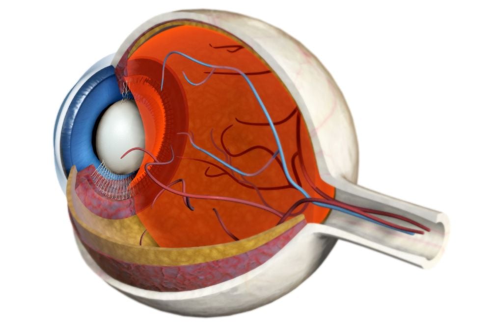
The retina is located at the back of the eyeball, on the wall, opposite the lens and pupil. Very thin, it only measures about 0.5 mm thick, about half the width of a sharp pencil point. Yet this tissue layer is crucial, enabling living things to see images. Upon entering through the eye’s lens, light hits the retina, converting it into an electrical signal. These accumulated signals then travel along the optic nerve to the brain, where they’re again transformed, into viewable images.
In terms of makeup, the retina has two parts, the macula and the peripheral retina, although other structures are vital to its function.
Macula
While responsible for sight, the retina’s yellow-pigmented center, the macula, allows for central vision. This is accomplished by the macula’s center, the fovea, which possesses the eye’s highest level of visual acuity, the ability to distinguish objects at a given distance. The fovea allows you to view straight-ahead, direct images and perform close-up activities, like cooking or driving.
Peripheral Retina
The peripheral retina allows you to see at the edges of your visual field. Also called peripheral, or side vision, it completes visual details and is what’s seen out of the corner of your eye.
Photoreceptor Cells
During the visual processing process, incoming light travels through multiple cell layers to reach the retina’s photoreceptors, light-sensitive cells. Upon making contact, light activates molecules called photopigments, which absorb light and improve visual processing. About 125 million photoreceptors, called rods and cones, work together to provide a clear, accurate picture, incorporating light, color, and fine details. Rods also help you see in dim light and at night, while cones, which make up most of your regular vision, process color. As humans have three different kinds of color-sensitive cone cells — red, green, and blue — we can see these three primary colors.
The outermost retinal layer is called the retinal pigment epithelium (RPE). This pigmented tissue layer contains many photoreceptors. This pigmented tissue layer contains the retina’s highest concentration of melanin, a natural eye pigment that absorbs damaging ultraviolet (UV) and blue light. The RPE also nourishes the retina and enables crucial metabolic processes.
Optic Nerve
Once activated, photopigment undergoes a chemical change, beginning the phototransduction process. This involves a signal being transmitted to bipolar cells, those connecting photoreceptors and ganglion cells, which exit the eye through an opening called the optic disk, a group of retinal tissue cells. An estimated 1.2 million retinal ganglion cell axons, or nerve fibers, then gather at the optic disc. Together, they form the optic nerve, located in the back of each eye, and directly connected to the brain.
The optic disc is responsible for transmitting electrical impulses from your eyes to your brain, which processes this information, enabling sight. Each eye’s optic nerve contains branches that travel to your brain or join with other fibers. When the two optic nerves cross, they form an “X”-shaped structure called the optic chiasm. Here, half of the left eye’s nerve fibers connect to the left side of the brain, and half of the right eye’s nerve fibers connect to the brain’s right side. As for the remaining nerve fibers, they combine. Signals from both eyes are received by the brain at the same time, forming a cohesive visual image, or binocular vision.
Educate Yourself on Retinal Makeup and Function
An essential structure, the retina affords the ability to see images. Understanding more about it can help you alert your doctor about any concerns that may arise. If you’d like a comprehensive retinal exam, or you have questions, please contact Vitreoretinal Consultants NYC to schedule a consultation.


